Drs. Joel L. Rosenlicht and Peter Vitruk discuss gingival hyperplasia and peri-implant pocket treatment using a CO2 laser
 Introduction
The term gingival hyperplasia refers to excess gingival tissue growth. It can be caused by numerous factors, most common being periodontal disease, poor oral hygiene, medications, smoking, and ill-fitting denture prostheses.1,2 The treatment of hyperplasia includes elimination of the causing factors and surgical removal of the lesion.1 If the cause persists, the tissue becomes more fibrous over time and can ulcerate and cause further pathology.
This article presents a case report of the surgical removal of gingival hyperplasia along with the sulcular debridement for peri-implantitis using the flexible fiber CO2 laser with a variety of dental and surgical laser handpieces.
Hyperplasia can be treated conservatively (by improving oral hygiene) or surgically (by scalpel, electrosurgery, carbon dioxide [CO2], Er:YAG, Nd:YAG, and diode lasers).1-3 The greatest disadvantage of the conventional scalpel is intraoperative hemorrhage and the need to suture the wound (patient discomfort).4 The CO2 laser in the case described in this article did not require sutures and allowed the surgeon to perform vestibuloplasty with a palatal mucosal graft during the same visit. Electrosurgery was not an option in the case described in this article because of the close proximity to the titanium implants.5 The CO2 laser is an excellent tool for removal of gingival hyperplasia because of its ability to induce hemostasis, speed, lack of the need for sutures, and excellent healing with reduced wound contraction without scar tissue resulting in healthy pliable tissue.6-8 In comparison with scalpel wounds, healing in the CO2 laser-treated wounds is characterized by a higher fibroblastic proliferation with young fibroblasts actively producing collagen. Only a small number of myofibroblasts (the cells responsible for wound contraction) are found in the CO2 laser-treated wounds compared with scalpel wounds.9,10 Thus, diminished wound contraction and reduced possibility of scar tissue formation are attributed to the insignificant amount of myofibroblasts.
 In addition, the CO2 laser can be effectively used for sulcular debridement due to its bactericidal properties along with the safety of the 10,600 nm wavelength around titanium implants.11-13
Soft tissue laser surgery
The CO2 laser is a “what you see is what you get” surgical soft tissue cutting laser with minimal collateral thermal effects sufficient for sealing blood vessels, lymphatics, and nerve endings; the surface bacteria are efficiently destroyed on incision/
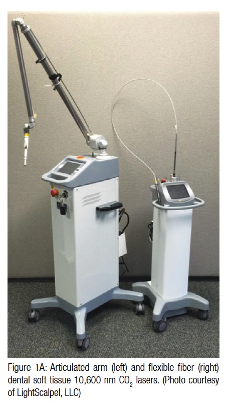
ablation margins. The current generation dental CO2 laser technology features a small foot-print, compact unit with flexible hollow fiber beam delivery (Figure 1A), and a variety of straight and angled handpieces (Figure 1B). The handpieces are pen-sized, disposable-free, autoclavable, and easily adapted to switching between (1) incision with coagulation, (2) superficial ablation with coagulation, or (3) coagulation modalities.
CO2 laser photo-thermal ablation and coagulation
Soft tissue photo-thermal ablation is a process of vaporization of intra- and extra-cellular water near the surface where the laser beam intensity is at its maximum (Figure 2). For a fixed laser beam diameter (or spot size), the volume of the tissue exposed to the laser beam is proportional to the optical penetration (i.e., absorption or Near-IR attenuation as defined earlier) depth. The shorter the penetration depth — the less energy is required to ablate the tissue. The longer the optical penetration depth — the greater the volume of irradiated tissue, and therefore, more energy is required to ablate the tissue within the irradiated volume of tissue. The 10,600 nm CO2 laser is highly energy efficient at ablating the soft tissue photo-thermally with very low ablation threshold intensities due to extremely small volume of irradiated tissue because of extremely short absorption depth around 15 µm.14
The coagulation zone is located immediately below the ablation zone (Figure 2). Coagulation occurs as a denaturation of soft tissue proteins that occurs in a 60°-100°C temperature range leading to a significant reduction in bleeding (and oozing of lymphatic liquids) on the margins of ablated tissue during laser ablation (and excision/incision) procedures. The coagulation depth value relative to the blood vessel diameter is an important measure of coagulation and hemostasis efficiency. For CO2 laser, its excellent coagulation efficiency is due to the close match between the photo-thermal coagulation depth of approximately 50-100 µm14 and oral soft tissue blood capillary diameters of approximately 20-40 µm.15
Thermal Relaxation Time
The rate of how fast the irradiated tissue diffuses the heat away is defined by Thermal Relaxation Time, which equals approximately 1.5 msec for 75% water rich soft tissue irradiated by 10,600 nm CO2 laser. Practical implications of the Thermal Relaxation Time concept are simple and yet very powerful for appropriate application of laser energy. The most efficient heating of the irradiated tissue takes place when laser pulse energy is high, and its duration is much shorter than TR. The most efficient cooling of the tissue adjacent to the ablated zone takes place if time duration between laser pulses is much greater than TR. Such laser pulsing is referred to as SuperPulse and is a must-have feature of any state-of-the-art soft tissue surgical CO2 laser that minimizes the depth of coagulation.
Laser beam spot size
Just as the sharpness of the steel blade defines the quality and the ease of the cut, the size of the laser beam focal spot defines the quality of the laser cut. The smaller (or sharper) the focal spot of the beam, the narrower and the deeper the incision. Just as a dull blade cannot produce a quality incision, an oversized laser beam spot cannot produce a precise and narrow incision. For a rapid switch from cutting to just photo-coagulation, the laser beam can be defocused either by selecting a larger spot size or by simply moving the handpiece away from the tissue by approximately 10 mm and “painting” the “bleeder” for enhanced hemostasis.
Laser power density and depth of ablation
Consider a steel blade: Regardless of how sharp the blade is, there will be no interaction between the blade and the tissue unless mechanical pressure is applied to the blade, forcing it through the tissue surface. For a laser scalpel, the power density of the focused laser beam is equivalent to the mechanical pressure that is applied to a cold steel blade: the greater the laser power density, the greater the depth and the rate of soft tissue removal.
 Controlling thermal effects
The SuperPulse setting (see preceding explanation) minimizes the amount of the heat transfer from the cutting/ablation zone to surrounding tissue; it results in minimal char on the margins of the cut, facilitating better healing and reduced postoperative scarring of the surgical wounds. For a superior hemostasis effect through photo-coagulation by laser light, turning the SuperPulse mode off is recommended; such a capacity of the CO2 laser is especially useful for procedures involving highly vascular tissues and patients with coagulation disorders or undergoing anticoagulant therapy.
Laser-implant interaction
In a recent study on CO2 laser removal of biofilms from implant surfaces,16 both titanium and titanium oxide (the most common implant materials) are reported to be un-
affected when treated by high power 10,600 nm CO2 laser. Figure 3 presents the absorption spectrum of the titanium surface. It illustrates that titanium implants, when treated by the CO2 laser, are affected (heated) approximately 4 times less than with diode and Nd:YAG wavelengths (circa 1,000 nm) and approximately 3 times less than with Erbium laser wavelengths (circa 3,000 nm).
A case report
Patient
The 66-year-old female patient presented for discomfort and pain, associated with her full arch mandibular implant-supported prosthesis. The patient had had the prosthesis for 10 years. Over that period of time, a significant amount of gingival hyperplasia developed around and between the implants along with a decrease in vestibular depth (Figure 4). Hyperplastic tissue around one of the implants was inflamed with 5 mm of pocket depth and some circumferential bone loss (cotton swab in Figure 4 points at the involved implant). It was decided to perform the following procedures utilizing the CO2 laser: (1) ablation of gingival hyperplasia, including the inflamed peri-implant tissue; (2) sulcus sterilization around the involved implant; and (3) vestibuloplasty with a palatal free gingival mucosal graft. Vestibular release and hemostasis for the palatal graft donor and recipient sites were done with the CO2 laser. The focus of this article is only on the reduction of gingival hyperplasia and sulcus sterilization immediately preceding the vestibuloplasty.


Laser equipment and settings
LightScalpel LS-2010 — the 20-watt carbon dioxide laser — (LightScalpel, LLC, Woodinville, Washington) was used with the following settings: 2-4 watts SuperPulse, F1 repeat pulse modes 3-8. Three handpieces were used during the procedure: a fixed spot tipless handpiece, an angled tipless handpiece, and an angled tip retainer handpiece with ceramic perio-focusing tip. All three of the handpieces had the laser beam spot size of 0.25 mm (the smallest available).
Surgical procedure
Topical anesthesia was applied to the site (Figure 4). Then local anesthesia was administered by infiltration (2% lidocaine with 1:100,000 epinephrine). Ablation of hyperplastic tissue was performed on the gingival tissue between the implants (Figure 5). The straight tipless handpiece was held at a 3-5-mm nozzle-to-tissue distance (Figure 5). The surgeon’s hand quickly moved in overlapping strokes as though “erasing” the excess tissue. Depending on the tissue amount and thickness, a number of passes were required to remove the hyperplasia from the gingiva.


After the hyperplastic tissue was vaporized in the spaces between implants, the inflamed tissue around the implant was ablated (Figure 6). The traces of carbonized ashes were rinsed off, and the surgical area was blotted off with damp gauze pad to ensure better penetration of the laser energy. In order to gain better access to the lingual part of the peri-implant tissue, the surgeon switched to the angled tipless handpiece (Figure 7). Once the inflamed tissue was removed and the surgical site rinsed off, touch-up ablation was performed to finish the hyperplastic tissue reduction between the implants (Figure 8).
For sulcular debridement, the surgeon switched to the angled handpiece with a ceramic periodontal tip (Figure 9). The laser power was reduced to 2 watts in the SuperPulse mode with repeat pulsing F1-3 mode. The periodontal tip was placed into the depth of the pocket and tracked slowly circumferentially around the implant to ablate the diseased tissue and eliminate the bacterial load within the pocket. Approximately 8-10 seconds were spent on both the buccal and lingual areas, with the tip smoothly gliding around the implant to ensure the laser energy reached all aspects of the peri-implant pocket (Figure 10).
After the peri-implant pocket treatment was finished, the pocket was thoroughly irrigated, and all residual intrasulcular material within the pocket was removed with implant curettes to make sure that all granulation tissue and residual epithelium had been eliminated.
The handpiece with the perio tip was then replaced by the tipless angled handpiece. Defocusing the laser was used to achieve hemostasis inside the peri-implant pocket where mild hemorrhage occurred from the mechanical debridement. Finally, the walls of the sulcus were smoothed out with the laser. The surgical area was rinsed off, and the procedure was completed (Figure 11 shows the surgical site immediately postoperatively). The patient was ready for the next stage of the surgery — vestibular tissue release and vestibuloplasty, which by doing so may contribute to the continued long-term success of this 10-year old prosthesis.
Postoperative assessment
The use of the laser for the tissue reduction, sulcular debridement, and vestibuloplasty effectively reduces postoperative pain and discomfort by the sealing off and ablation of the exposed tissue. This reduces postoperative complaints of swelling and bleeding and minimizes the need for excessive post-surgical analgesia. The patient’s prosthesis was immediately reinserted following the procedure and function restored.
Follow-up exam
The patient was seen 1 week after the surgery. Healing was uneventful, and the patient was pleased with the outcome. Typically, the tissue reduction site is well healed within 7-10 days and the vestibuloplasty site within 2-3 weeks.
Conclusion
The CO2 laser was efficiently used for the hyperplastic gingival ablation and sulcular debridement. The ability of the CO2 laser to achieve hemostasis proved especially important for the procedures described in this article for the following reasons:
The laser maintained the dry bloodless operatory field with excellent visibility.
Ability to see ensured precise tissue removal with very little collateral damage to the surrounding un-
affected tissues.
No sutures were required reducing overall surgery time.
Patients report less post-op pain and swelling, thus less analgesia is necessary.
With bactericidal effects, many at-risk implants can be treated early, before too much of the supporting bone is lost.
Acknowledgments
The authors greatly appreciate the support and contribution from Anna Glazkova, PhD, in preparing this material for publication.
This article is sponsored by LightScalpel, LLC.
(www.LightScalpel.com, 1-866-589-2722)
 Joel L. Rosenlicht, DMD, an oral and maxillofacial surgeon, is past president of the American Academy of Implant Dentistry (AAID) and past president of the American College of Oral and Maxillofacial Surgeons (ACOMS). He is a Diplomate of the American Board of Oral and Maxillofacial Surgery (ABOMS) and of the American Board of Implant Dentistry (ABOI).
 Peter Vitruk, PhD, MInstP, CPhys, is founder of LightScalpel, LLC. He is a member of the The Institute of Physics and of the Science and Research Committee, Academy of Laser Dentistry. He is also on the faculty of the California Implant Institute and Global Laser Oral Health, LLC. Dr. Vitruk can be reached at 1-866-589-2722 or pvitruk@LightScalpel.com.
-
Convissar RA, Sawisch TJ, Strauss RA. Laser-enhanced removable prosthetic reconstruction. In: Convissar RA. Principles and Practices of Laser Dentistry. St. Louis, MO: Mosby;2011:93-113.
-
de Arruda Paes-Junior TJ, Cavalcanti SC, Nascimento DF, Saavedra Gde S, Kimpara ET, Borges AL, Niccoli-Filho W, Komori PC. CO2 Laser Surgery and Prosthetic Management for the Treatment of Epulis Fissuratum. ISRN Dent. 2011;2011:282361. doi: 10.5402/2011/282361.
-
Namour S. Atlas of Current Oral Laser Surgery. Boca Raton, FL: Universal Publishers. 2011;139-171.
-
Sawisch TJ. Oral surgery for the general practitioner: ablation/ vaporization techniques and procedures – clinical scenarios. In: Convissar RA. Principles and Practices of Laser Dentistry. St. Louis, MO: Mosby; 2011:93-113.
-
Convissar RA, Gharemani EH. Laser treatment as an adjunct to removable prosthetic care. Gen Dent. 1995;43(4):336-341. Points out that the CO2 laser, unlike the conventional scalpel, allows gradually, cell layer at a time, removing precisely the right amount of tissue.
-
Zeinoun T, Nammour S, Dourov N, Aftimos G, Luomanen M. Myofibroblasts in healing laser excision wounds. Lasers Surg Med. 2001;28(1):74-79.
-
Strauss RA, Fallon SD. Lasers in contemporary oral and maxillofacial surgery. Dent Clin North Am. 2004;48(4):861-888.
-
Wlodawsky RN, Strauss RA. Intraoral laser surgery. Oral Maxillofac Surg Clin North Am. 2004;16(2):149-163.
-
Grbavac RA, Veeck EB, Bernard JP, Ramalho LM, Pinheiro AL. Effects of laser therapy in CO2 laser wounds in rats. Photomed Laser Surg. 2006;24(3):389-396.
-
de Freitas AC, Pinheiro AL, de Oliveira MG, Ramalho LM. Assessment of the behavior of myofibroblasts on scalpel and CO2 laser wounds: an immunohistochemical study in rats. J Clin Laser Med Surg. 2002;20(4):221-225.
-
Kato T, Kusakari H, Hoshino E. Bactericidal efficacy of carbon dioxide laser against bacteria-contaminated titanium implant and subsequent cellular adhesion to irradiated area. Lasers Surg Med. 1998;23(5):299-309.
-
Deppe H, Horch HH, Henke J, Donath K. Peri-implant care of ailing implants with the carbon dioxide laser. Int J Oral Maxillofac Implants. 2001;16(5):659-667.
-
Deppe H, Horch HH, Greim H, Brill T, Wagenpfeil S, Donath K. Peri-implant care with the CO2 laser: In vitro and in vivo results. Med Laser Application. 2005;20(1):61-70.
-
Vitruk P. Oral soft tissue laser ablative & coagulative efficiencies spectra. Implant Practice US, 2014;7(6):22-27.
-
Yoshida S, Noguchi K, Imura K, Miwa Y, Sunohara M, Sato I. A morphological study of the blood vessels associated with periodontal probing depth in human gingival tissue. Okajimas Folia Anat Jpn. 2011;88(3):103-109.
-
Cobb CM, Vitruk P. Microbial Decontamination of Three Different Implant Surfaces Using a SuperPulse CO2 (10,600 nm) Laser: An In Vitro Study. Presented at the Academy of Laser Dentistry Meeting. Palm Springs, February 5-7, 2015.
-
Wolfe WL, Zissis GJ. The Infrared Handbook. Washington DC: Office of Naval Research, NAVY, 1985;7-81.
Stay Relevant With Implant Practice US
Join our email list for CE courses and webinars, articles and mores

