Dr. Randolph Resnik finds that 3D cone beam imaging shows incidental findings that become integral in the planning of a successful course of treatment
In an article in Implant Practice US, entitled “CBCT and Implants: the new era in treatment planning and diagnosis” (October 2013, Vol. 6, No. 5), I discussed 3D cone beam imaging (CBCT) and its clinical benefits for treatment planning and placement of dental implants. The information obtained from a scan of the patient’s 3-dimensional anatomy may significantly impact the predictability of the implant procedure.
Information about anatomical landmarks, such as the inferior alveolar nerve, maxillary sinus, nasal cavity, and mandibular lingual concavities, help the implant dentist to prudently plan treatment. In addition to these vital structures, there are anatomic variants that may be visible on 3D scans that may not be evident with 2D radiography. Recent studies show that the average scan contains approximately 3.2 incidental findings per CBCT scan.1 Thus, the following case study reveals the importance of identifying and understanding “incidental” findings, which may impact future dental implant planning.
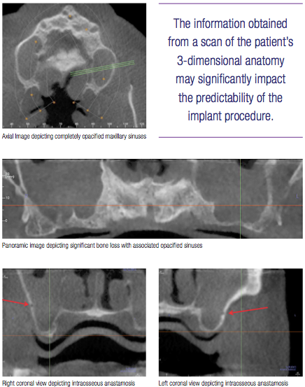
A 55-year-old male presented to my office for evaluation and treatment alternatives for an ill-fitting maxillary complete denture. His medical history was unremarkable with no known allergies. My office obtained an i-CAT® FLX (Imaging Sciences International) scan for evaluation of future implants. Two incidental findings were noted: (1) Both maxillary sinuses were completely radiopaque. (2) The lateral wall of the maxillary sinus depicted a large intraosseous anastomosis.
The presence of bilateral radiopaque sinuses is consistent with significant maxillary sinus disease; however, the patient was completely asymptomatic. This presented the opportunity for me to refer the patient to an otolaryngologist (ENT). Besides the obvious advantages to treating sinus pathology before any implant or bone grafting procedures involving the maxillary sinus, there exists an additional benefit. By having the patient treated and cleared for future procedures in the maxillary sinus with an ENT, the implant dentist has the ability to form a relationship with an ENT physician that will be crucial if postoperative complications develop.
In this particular case, the ENT performed a procedure termed functional endoscopic sinus surgery (FESS), which entails placing an endoscope through the maxillary sinus ostium. With this treatment, the ostium is opened, and the sinus is cleaned to reduce the pathology present. After a course of antibiotics, the patient was cleared for future bone grafting and dental implant treatment.
The other finding, the intraosseous anastomosis, presented as a large radiolucent concavity in the lateral wall of the maxillary sinus. This particular anatomic variant is a relatively new finding in the dental implant literature, which represents an anastomosis of posterior superior artery and the infraorbital artery. So, why is this important? In a lateral sinus graft, a window is made on the lateral aspect of the maxillary sinus where this anastomosis is present. Preoperatively seeing this variant on the CBCT scan allows the clinician to prepare for possible excessive bleeding during this procedure. In some instances, bleeding from this area is very significant.
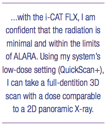
Having an i-CAT scan allows me to view all areas of concern. And with the i-CAT FLX, I am confident that the radiation is minimal and within the limits of ALARA. Using my system’s low-dose setting (QuickScan+), I can take a full-dentition 3D scan with a dose comparable to a 2D panoramic X-ray.2
As a result, it is well within a safe range if I need to repeat a scan after an implant is placed or to evaluate healing of a bone graft.
 In this case study, the importance of a CBCT scan is easily seen. Without a radiographic exam of this type, the patient’s pathologic maxillary sinuses may have gone undetected, thus placing the patient at an increased morbidity for procedures in this area. Additionally, the patient was able to be treated with sinus bone grafting and future implants for a fixed maxillary prosthesis.
Besides performing a quality implant procedure, CBCT is integral in helping my patients. Whether working with an ENT, or sharing my scans with a referring dentist, I would not want to work without 3D imaging, its details, and capacity for low radiation dose. This patient now trusts that I am interested in more than his teeth — his quality of life has improved, and now I am more ready to improve the quality of his dentition through implants as well.
References
1.Price JB, Thaw KL, Tyndall DA, Ludlow JB, Padilla RJ. Incidental findings from cone beam computed tomography of the maxillofacial region: a descriptive retrospective study. ClinOralImplantsRes. 2012;23(11):1261-1268.
2.Ludlow JB, Walker C. Assessment of phantom dosimetry and image quality of i-CAT FLX cone-beam computed tomography. AmJOrthodDentofacialOrthop. 2013;144(6):802-817.
Stay Relevant with Implant Practice US
Join our email list for CE courses and webinars, articles and mores
Read our following terms and conditions before subscribing.
